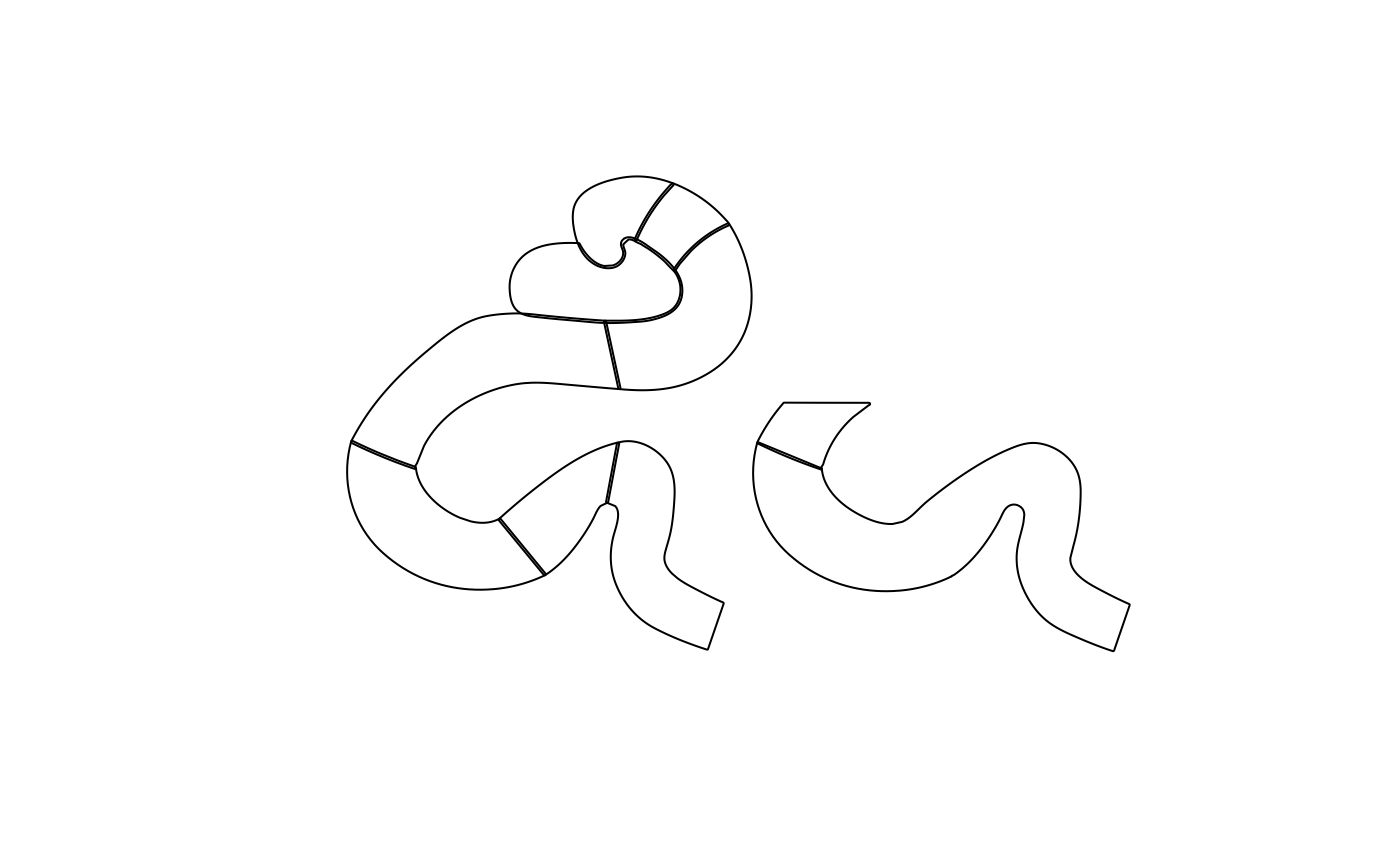
Medial temporal lobe atlas for T2-weighted anatomy Anika Wuestefeld
mtl_t2.RdA two-dimensional medial temporal lobe atlas imported from a labelled SVG drawing. The atlas represents medial temporal cortical regions and hippocampal subfields.
Format
An sf object with 9 rows and 2 columns:
- region
Character anatomical region label. Labels are
"ERC"(entorhinal cortex),"BA35"and"BA36"(Brodmann areas 35 and 36),"SUB"(subiculum),"CA1","CA2", and"CA3"(cornu ammonis subfields),"PHC"(parahippocampal cortex), and"CA3-2"(the second disconnected CA3 component in the drawing).- geometry
Simple-feature polygon or multipolygon geometry in the coordinate system of the source SVG drawing. The atlas has no coordinate reference system.
Details
This is a schematic plotting atlas, not a spatially registered
neuroimaging atlas. It was created from a labelled SVG drawing with
build_atlas_svg().
Examples
data(mtl_t2)
plot(sf::st_geometry(mtl_t2))